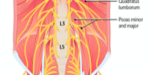
Anatomy of the Posterior Abdominal Wall


It might not be the flashiest anatomical structure, but if you want to stand upright, and keep your retroperitoneal organs (like your kidneys) in place, the posterior abdominal wall is pretty important. Located at the back of the body, bounded by the lateral abdominal walls and the posterior parietal peritoneum, the posterior abdominal wall is a complex combination of muscles, bones, nerves, and vessels that provides structural support for the body and for the organs of the retroperitoneal space.
After listening to this AudioBrick, you should be able to:
- Describe the structure and relationships of the posterior abdominal wall, including the aorta (including collateral channels), inferior vena cava (including collateral tributaries), lymphatics, muscles (psoas major, quadratus lumborum), sympathetic chain, and lumbar plexus.
- Describe the relationship of the kidneys in the retroperitoneal space as it relates to posterior abdominal wall anatomy.
You can also check out the original brick from our Cardiovascular collection, which is available for free.
Learn more about Rx Bricks by signing up for a free USMLE-Rx account: www.usmle-rx.com
You will get 5 days of full access to our Rx360+ program, including nearly 800 Rx Bricks. After the 5-day period, you will still be able to access over 150 free bricks, including the entire collections for General Microbiology and Cellular and Molecular Biology.
***
If you enjoyed this episode, we’d love for you to leave a review on Apple Podcasts. It helps with our visibility, and the more med students (or future med students) listen to the podcast, the more we can provide to the future physicians of the world.
Follow USMLE-Rx at:
Facebook: www.facebook.com/usmlerx
Blog: www.firstaidteam.com
Twitter: https://twitter.com/firstaidteam
Instagram: https://www.instagram.com/firstaidteam/
YouTube: www.youtube.com/USMLERX
Learn how you can access over 150 of our bricks for FREE: https://usmlerx.wpengine.com/free-bricks/
