STEP 1 Question of the Month (February)

Conquer STEP 1, One Question at a Time! The answer is explained below.
A 36-year-old nulliparous woman comes to her primary care physician because of breast pain for several days. She says she has felt lumps in her breasts previously. These lumps worsen the week before and around the time of her period but subside afterward. Breast examination shows several small, mobile lumps on both breasts. There is no axillary lymphadenopathy.
Which of the following would a biopsy specimen of the breast lumps most likely reveal?
A) Central necrosis
B) Increase in number of acini and intralobular fibrosis
C) Large cells with clear “halos”
D) Lymphatic involvement
E) Sheets of pleomorphic cells infiltrating adjacent stroma
Check the answer below 👇
.
.
.
.
.
.
.
.
The correct answer is: B) Increase in number of acini and intralobular fibrosis.
High-Yield Summary
- Bilateral breast pain and cystic lumps without skin changes, nipple discharge, or axillary lymphadenopathy that worsen before menses and with caffeine intake is a typical presentation of fibrocystic breast changes.
- Histologically, fibrocystic breast changes appear as fibrous tissue, cysts, and possible proliferation of acini.
Step 1: Disease Diagnosis
This patient presents with cyclic, premenstrual breast pain with no additional symptoms and multiple bilateral, small, mobile lumps; no skin changes; and no axillary lymphadenopathy. This is a classic presentation of fibrocystic changes of the breast, which typically fluctuate in relation to the hormonal effects of estrogen and may be aggravated by caffeine intake.
Step 2: Normal Structure/Function
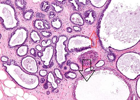
Fibrocystic disease is a benign condition that appears as fibrous tissue, cysts, and possible proliferation of acini on histologic examination. It manifests as diffuse breast pain and multiple bilateral cystic masses, typically in the outer quadrants. It is one of the most common causes of breast lumps in women, from the onset of menses to the onset of menopause. Fibrocystic breast tissue histologically appears as fibrous stroma (red arrow) and cysts (black arrow). There may be focal proliferative changes in some cases with an increase in the number of acini (terminal breast unit) per lobe.
The other choices are incorrect:
- Central necrosis is characteristic of comedocarcinoma, a subtype of ductal carcinoma in situ that presents as a unilateral, palpable mass with nipple discharge.
- Large cells with clear “halos” are seen in Paget disease, which occurs unilaterally with eczematous skin findings and is associated with underlying ductal carcinomas.
- Lymphatic involvement of breast tissue is indicative of inflammatory carcinoma, which presents with peau d’orange appearance of the skin and erythema and swelling.
- Sheets of pleomorphic cells infiltrating adjacent stroma are seen in invasive ductal carcinoma, which presents in older women as fixed, irregular masses with skin changes such as retraction, dimpling, and/or unilateral bloody nipple discharge.
Don’t Wait – Sign Up Now!
When you sign up for a free account, you’ll have access to Rx360+ for the first five days after sign-up. Take advantage of that time to test-drive the most comprehensive study system on the market and see the difference we can make in your studying.